| Veterinary
Histology UFF Department of Morphology - Biomedic Institute LaBEc - Laboratory of Cellular and Extracellular Biomorphology |
|||
Veterinary
Histology Atlas |
|||
Digestive
System |
|||||
Funçtion Structure • Hollow Tube composed of a lumen, whose diameter is variable, surrounded by a wall formed by four distinct layers: Mucosa |
|||||
| Muscularis
Mucosae • Separates the Mucosa from the Submucosa • Normally composed of two thin sub-layers of smooth muscle cells |
|||||
Submucosa |
|||||
Muscularis
Layers |
|||||
| • Between these two sub-layers we find the Myenteric Nervous Plexus or Auerbach’s Plexus | |||||
| |
Serosa |
||||
|
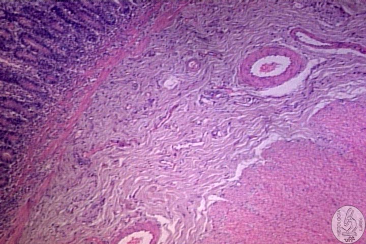
Adventitia • Thin Layer of Loose Connective Tissue, rich in blood vessels, lymphatic vessels and Adipose tissue • Does not possess Mesothelium • Found where the Digestive Organ is joined with other Organs or structures. |
||||
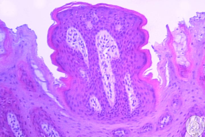
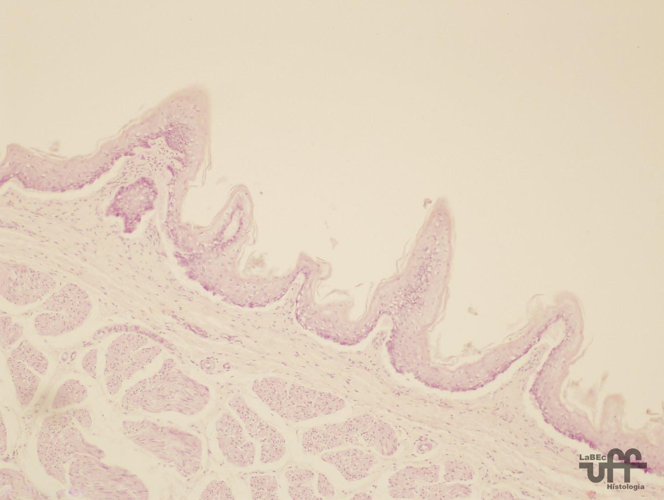
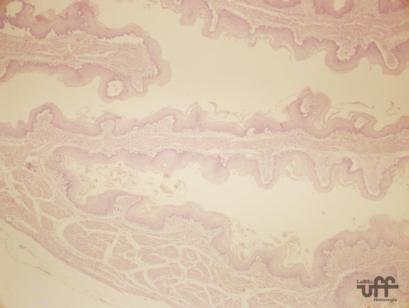
Constituition Tongue |
|||||
| •
Taste Buds: Specialized structures that contain taste bud cells,
substance detectors capable of drawing the flavor. |
|||||
| Papillae | |||||
|
|||||
| I
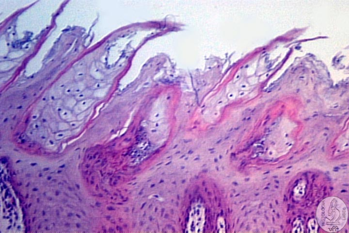
- Filiform • Have an elongated cone shape • Numerous • Present in the entire dorsal surface of the tongue • Mechanical action, do not possess taste buds |
II
- Fungiform • Resembles a mushroom, with a strait base and a more dilated and smooth superior surface portion • Irregularly distributed between the filiform papillae • Possess few taste buds |
||||
 |
 |
||||
| III
- Foliate • Poorly developed in humans, characteristic in rabbits • Consists on two or more parallel folds separated by grooves • Located on the dorsolateral surface of the tongue • Contain many taste buds |
IV
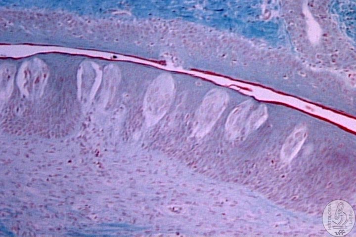
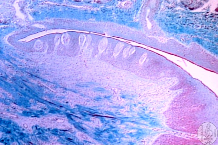
- Circumvallate • Great circular structures, whose flat surface extends itself to the top of other papillae • A Deep Depression surrounds each papilla • Present glands of von Ebner -Numerous Serous glands located in the fossa of each papilla -Continuous flow of liquid over the taste buds, important for the removal of food particles -Secrete lipase that prevents the formation of a hydrophobic layer over the taste buds • Distributed in the V-shaped boundary, in the posterior section of the tongue |
||||
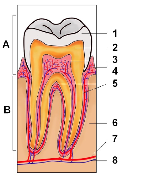
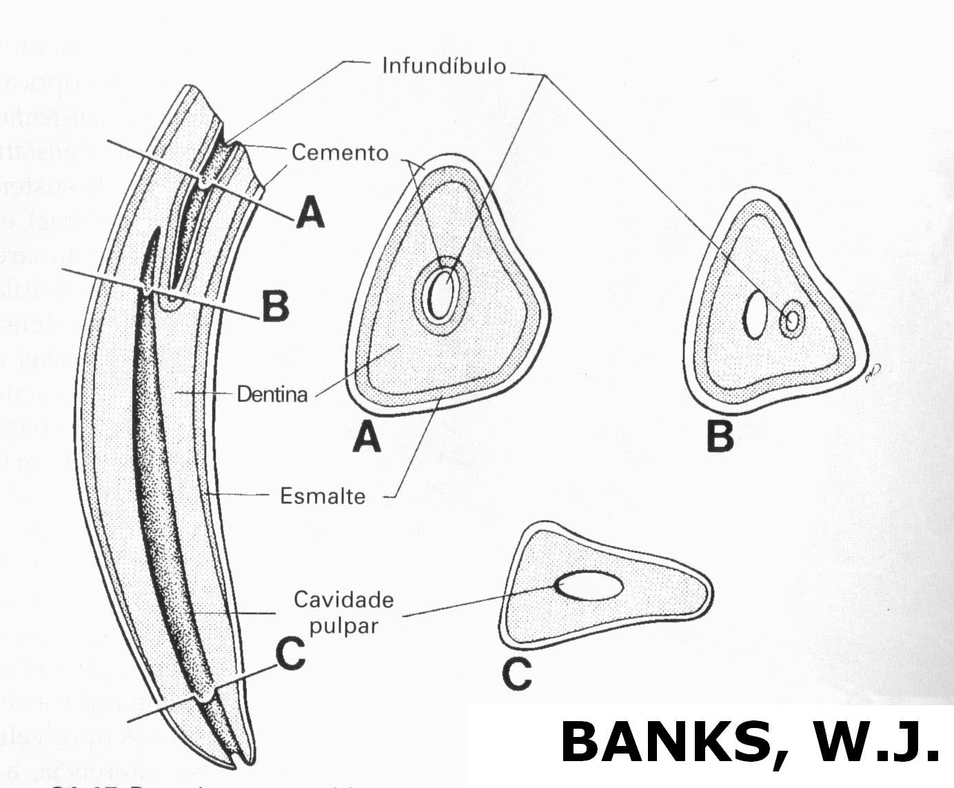
Teeth and Associated Structures Classification |
|||||
| |
Brachydonts |
||||
|
|
Hypsodonts |
||||
| Teeth Components | |||||
| Enamel |
 |
||||
•
The Organic Matrix is synthesized by ameloblasts
|
|||||
Dentin
|
|||||
Dental
Pulp |
|||||
| Periodontal Components | |||||
| Cementum
|
 |
||||
Periodontal
Ligament Alveolar
Bone Gingiva |
|||||
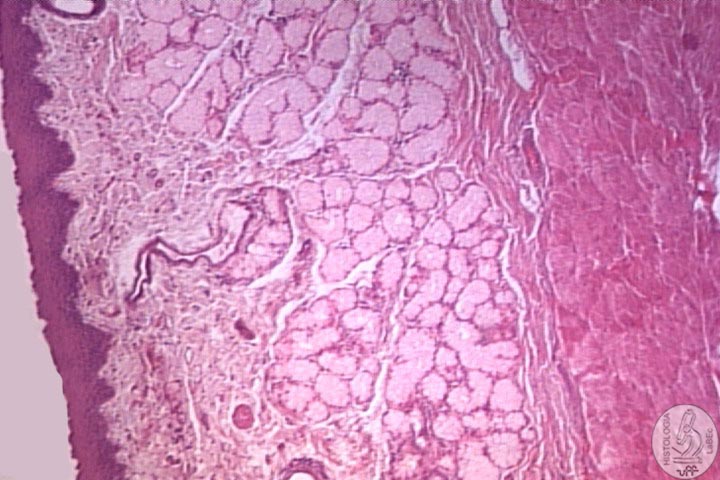
Oropharynx |
|||||
Esophagus |
|||||
| •
Muscular tube whose function is to transport the food from the mouth
to the stomach • Lined by Keratinized(or not)Stratified Squamous Epithelium, depends on the species • Typical Lamina Propria • Muscularis Mucosae formed by well fragmented smooth muscle fibers |
|||||
| •
Typical Submucosa containing mucous secreting esophageal glands,
that facilitate the transport of food and protect the mucosa • Muscularis Layer: Fibers are in different directions to facilitate the peristaltic movements -Upper Third: Only Striated Skeletal Muscle Fibers -Middle Third: Mix of Smooth muscle with Striated Skeletal muscle -Inferior Third: Only Smooth Muscle Fibers • Present Adventitia for there is a union of the esophagus with the trachea • Present Serosa in the Esophagus Region that is in the Peritoneal Cavity |
|||||
| Curiosities • The facility of ruminants and dogs to vomit and/or regurgitate is linked to the distribution of Striated Skeletal Muscle • Fecal Emesis and Regurgitation are not physiological events in dogs • Regurgitation is a physiological event in Ruminants • Even though equines posses Striated Skeletal Muscle in 2/3 of their esophagus, their vomit is very rare, because the caudal esophageal sphincter has enough tonus to remain closed during gastric dilation to a point where a stomach rupture occurs without the occurrence of vomit. |
|||||
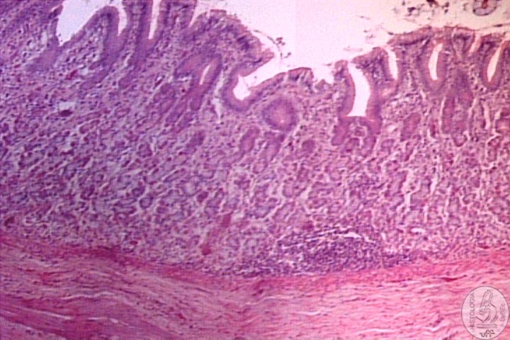
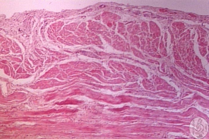
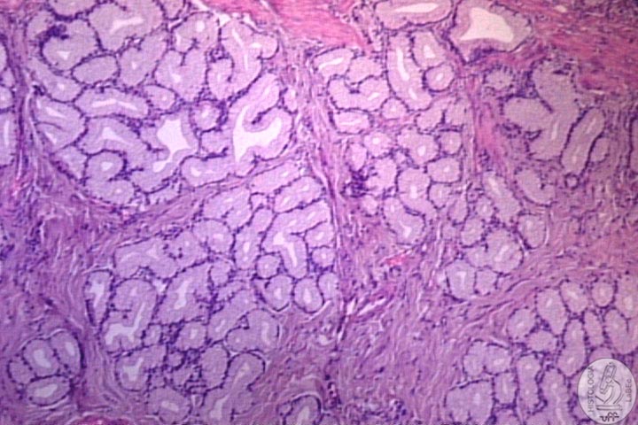
Stomach Division Monogastric Animals Structure |
|||||
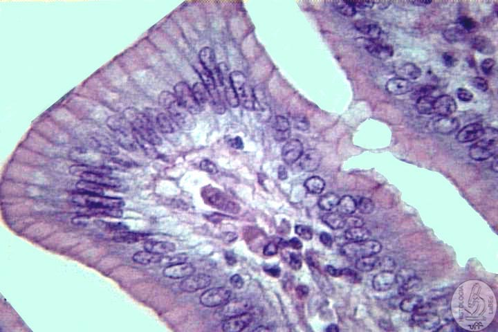
| •
Lined by Mucous Simple Columnar Epithelium(all of the cells secrete
an alkaline mucus) • The mucous forms a thick layer of gel that protects the cells from the stomach’s acidity • Occlusive Junctions between superficial cells and gastric pits form a protective barrier against the acid • HCl, Pepsins and Lipases are considered endogenous factors that damages the mucosa |
|||||
| •
Gastric Pits: -Epithelium that suffers invagination towards the lamina propria -In these pits characteristic glands of each region debouch secretion |
|||||
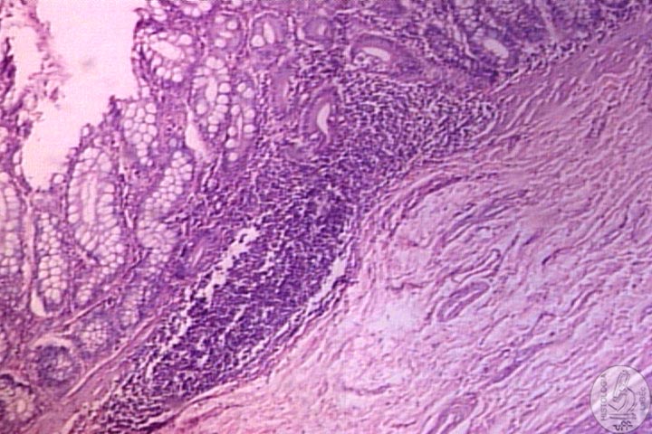
| •
Typical Lamina Propria containing smooth muscle cells and lymphoid
cells • Submucosa with dense connective tissue containing blood and lymphatic vessels |
|||||
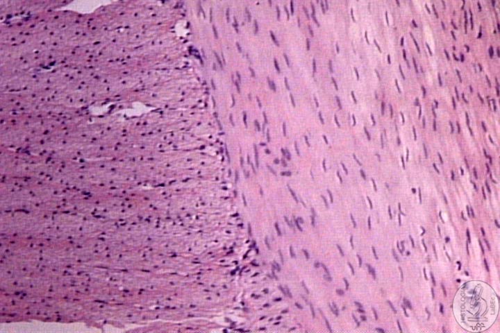
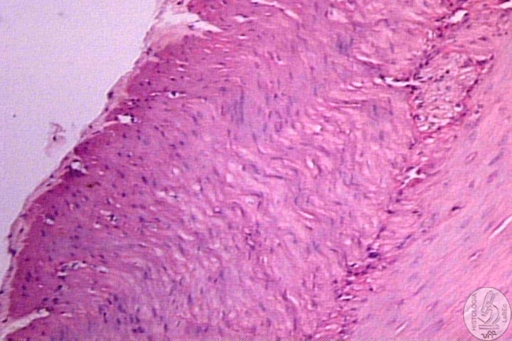
| •
Muscularis Layer contains Smooth Muscle Fibers oriented in three
main directions: -Inner Oblique -Middle Circular -Outer Longitudinal • Present a Thin Serosa |
|||||
Cell Types Stem-cells Mucous
Neck Cells |
|||||
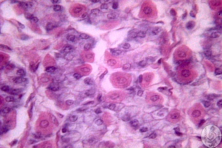
| Parietal
Cells • Round or Pyramid-shaped Cells with a central spherical nucleus, cytoplasm intensely acidophilic • Produce H+ and Cl- • Gastrin and Histamine are potent stimulators of HCl production • Gastrina possesses a trophic effect in the gastric mucosa, stimulating its growth • Are mainly present in the upper half of the Gastric Glands, scarce in its base |
|||||
| Zymogenic
Cells Enteroendocrine
Cells |
|||||
Histological Regions |
|||||
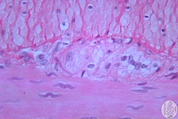
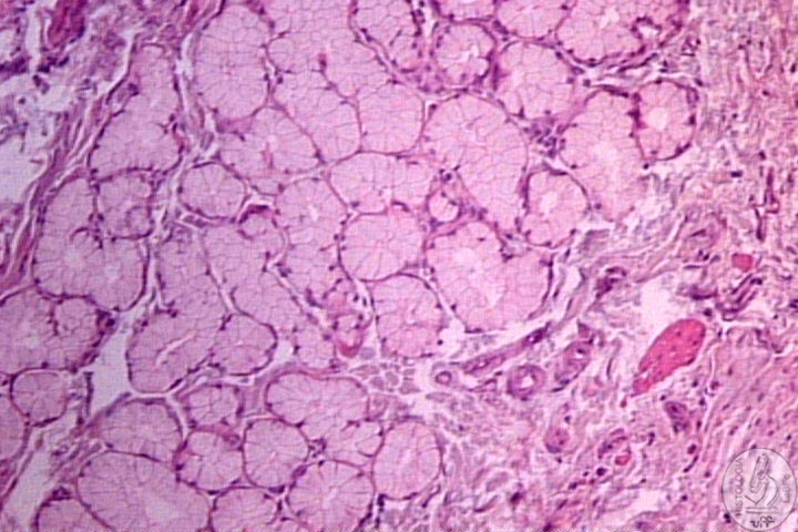
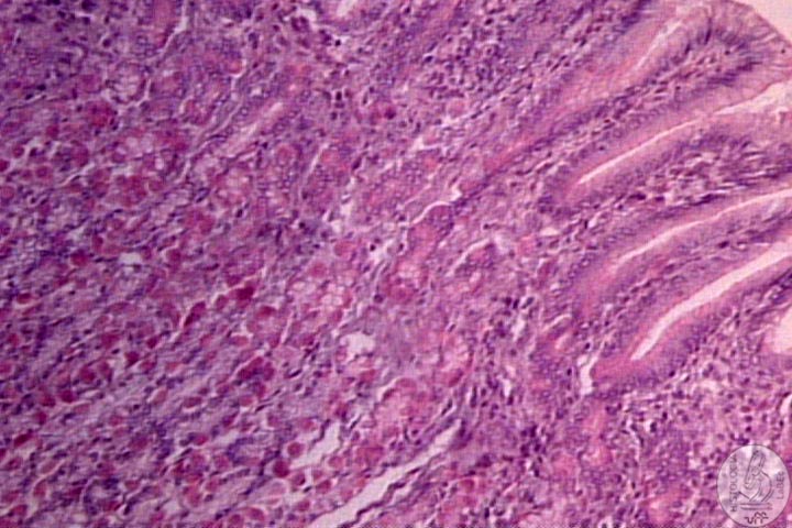
| Cardia • Transition between esophagus and stomach • Transition from Stratified Squamous Epithelium of the Esophagus to Mucous Simple Columnar of the Stomach • Submucosa contains cardiac glands that produce mucous and lysozyme(antibacterial) • Present few Parietal Cells |
|||||
| |
Fundus
(Corpus e Fundus are Histologically identical) |
||||
| Pylorus • Lined by Mucous Simple Columnar Epithelium • Possess Deep Gastric Pits where Pyloric glands open(mucus and lysozyme) • Present Enteroendocrine cells that secrete gastrin intercalated by Mucous Cells • The Middle Muscle Layer is thicker so to form a pyloric sphincter |
|||||
Polygastric Animals Forestomach |
|||||
| Rumen
(Paunçh) • Small Rumen Papillae • Do not possess Muscularis Mucosae (lamina propria and connective tissue of the submucosa are fused) • Have a mechanical function • Curiosities: -Produce Volatile Fatty Acids -Depend on protozoa and bacteria to digest -Timpanism -Gases |
|||||
| Reticulum
(Bonnet) • Reticulum' folds with a honeycomb aspect and spear-shaped • Muscularis Mucosae projects itself a little, present in the apex of the papilla |
|||||
| Omasum
(Manyplies) •Omasum' folds • Omasum' Folds with smaller papillae • Muscularis Mucosae projects itself a lot, forming an axis of smooth muscle in all of its extension. There is also a layer of smooth muscle of inner circular. • Retains food, it’s the coarsest papillae |
|||||
| True
Stomach-Obomasum(rennet-bag) • Same conformation as the common stomach • Regions: Nonglandular, Cardia, Fundus and Pylorus |
|||||
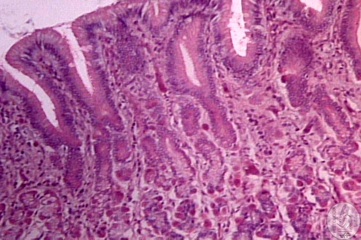
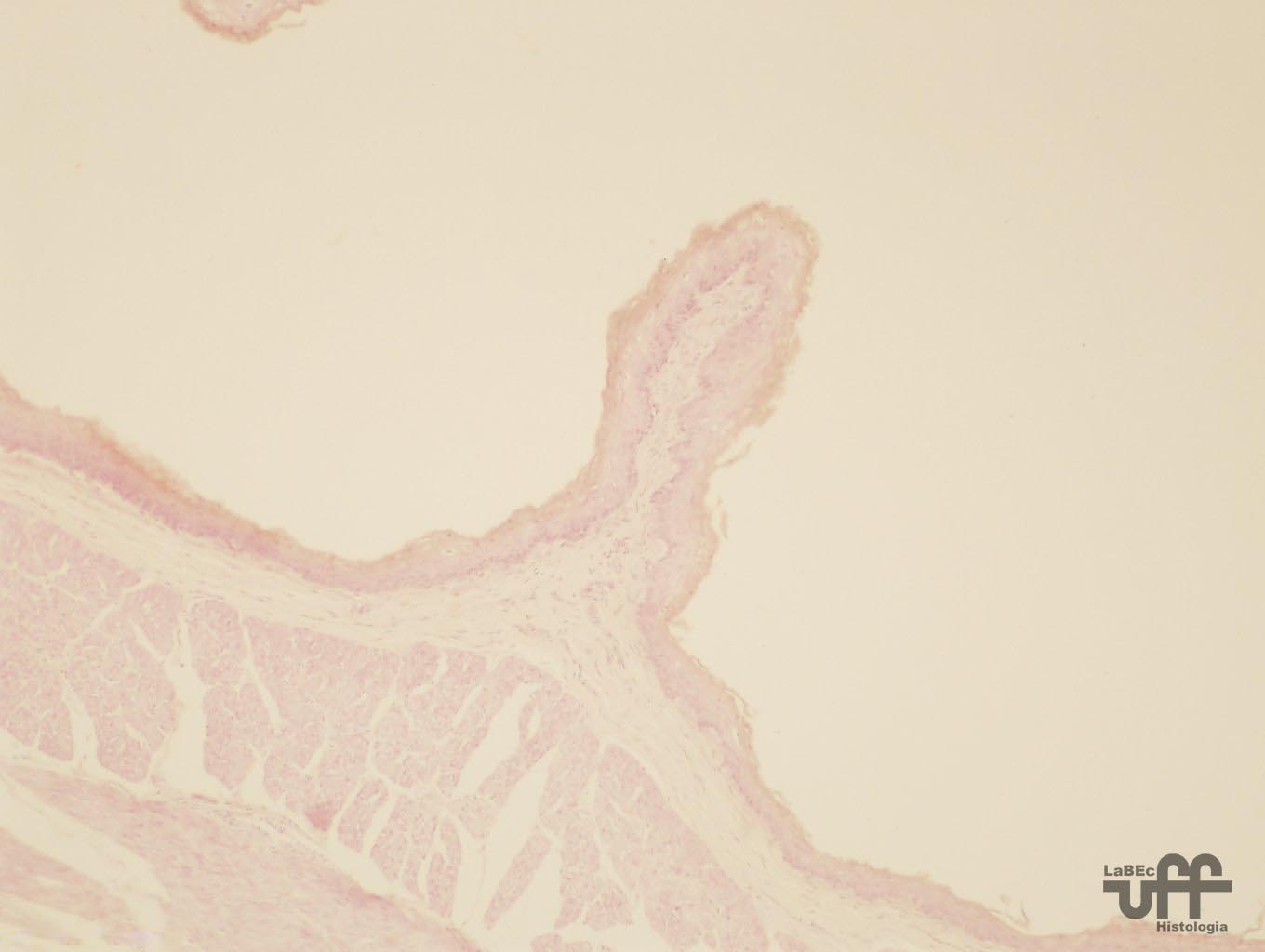
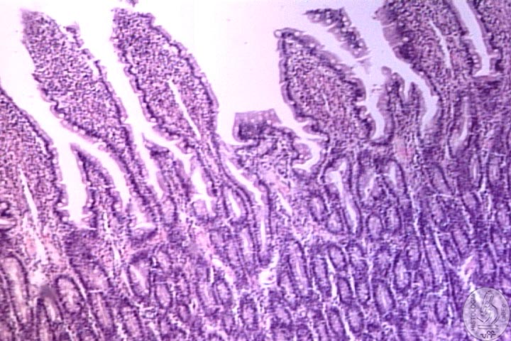
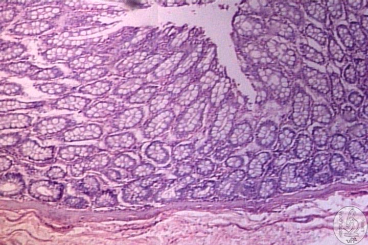
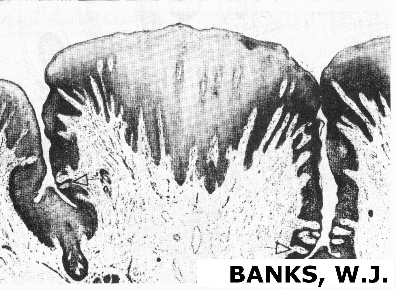
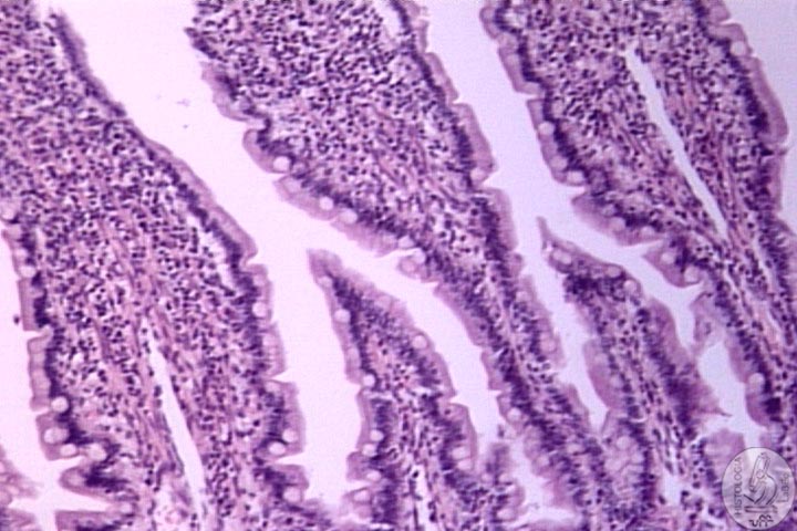
| Small
Intestine • Final part of the food digestion, nutrients absorption and endocrine secretion • Possess many modifications that enhance the contact surface and consequently the absorption: -Long length(5 meters) -Folds in Mucosa and Submucosa -Villi and Microvilli -Absorptive Cells(Enterocytes) *Type of epithelial cell of the superficial layer of the small and large intestines *These cells break-down molecules and transport them inside the tissue |
|||||
| Structure | |||||
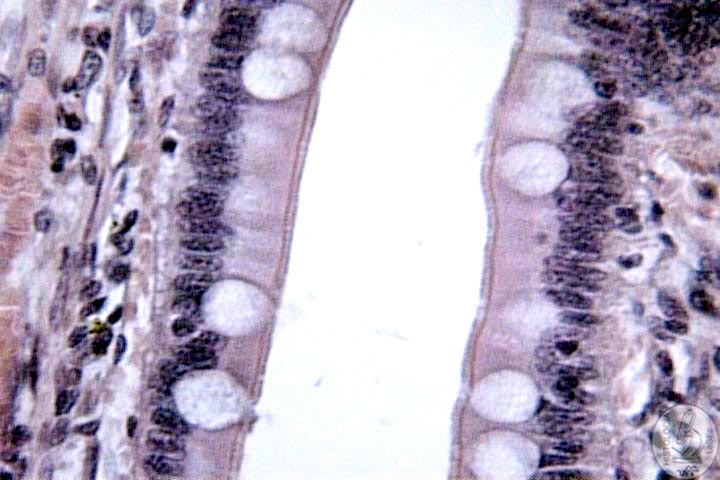
| •
Lined
by Simple Columnar Epithelium with a Striated Border and Goblet
cells(vary according to the portion of the intestine) |
|||||
| |
•
Present intestinal villi: Elongated Finger-like structures |
||||
Tipos Celulares Stem-Cells Absorptive
Cells (Enterocytes) Goblet
Cells Paneth
Cells M
Cells |
|||||
Division |
|||||
 |
Duodenum • Tube of approximately 2 to 3 centimeters in diameter that connects the stomach to the small intestine • Villi with a leaf-like aspect, close to the ileum they acquire a finger-like shape • Lined by Simple Columnar Epithelium with Striated Border and some RARE Goblet Cells • Present many Absorptive Cells and Paneth Cells • M cells and Goblet cells are characteristic of this region • Typical Lamina Propria • Muscularis Mucosa without any peculiarities |
||||
|
|
• Submucosa containing duodenal glands or Brünner Glands that produce mucus -This mucus protects the mucosa against the acidity of the gastric acid and neutralizes the pH of the chime -These glands open at the bottom of the Crypts of Lieberkühn • Muscularis Layer with an Inner circular and Outer Longitudinal layer and present Myenteric Plexus of Auerbach • Typical Serosa |
||||
Jejunum
|
 |
||||
 |
Ileum |
||||
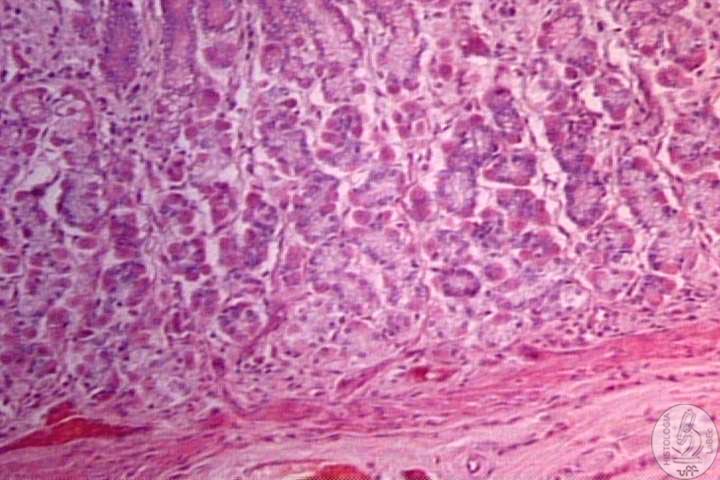
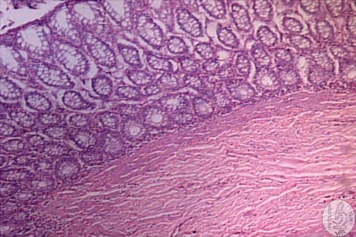
Large Intestine |
|||||
| |
•
Consists of a Mucosa Membrane filled with Goblet Cells, no Folds
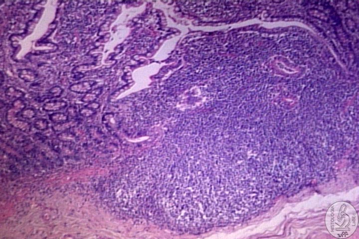
or Villi • Functions are water absorption, formation of feces and production of mucus • Lined by Simple Columnar Epithelium with a Striated Border(irregular and spaced) and MANY Goblet Cells • Present many Absorptive Cells and Goblet Cells, and some Enteroendocrine Cells • M Cells and Paneth Cells are not typical of this region • Lamina Propria with many lymphoid cells and nodules that frequently reach the submucosa • Muscularis Mucosae without any peculiarities • Typical Submucosa with lymphoid nodules • Muscularis Externa formed by three thick laminae(taenia coli) • Typical Serosa only until the neck after that it continues as an adventitia |
||||
|
|||||
Rectum |
|||||
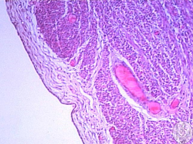
| Annus • The annus is the orifice at the end of the large intestine where the feces and intestinal gases are eliminated. • Transition from the Simple Columnar Epithelium to the Stratified Squamous, typical of the tegument • Lamina Propria containing a plexus of large veins • Adventitia |
|||||