| Veterinary
Histology UFF Department of Morphology - Biomedic Institute LaBEc - Laboratory of Cellular and Extracellular Biomorphology |
|||
Veterinary
Histology Atlas |
|||
Connective
Tissue Proper |
|
General Characteristics •
Heterogeneous Cell Population Functions •
Support (Physical Support) |
|
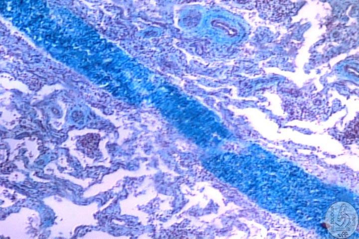
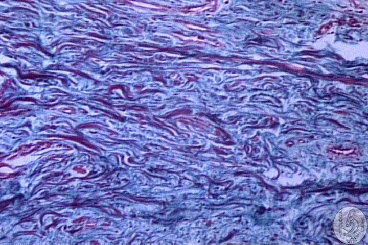
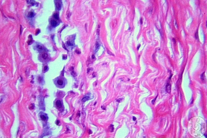
Classification Dense Connective Tissue: Few Cells, there are mostly fibers and extracellular matrix |
|
Regular:
Fibers organized towards the same direction |
|
Irregular:
Fibers don’t have a specific organization |
|
Loose
Connective Tissue:
Present
Fibers and Extracellular Matrix with a predominance of cells |
|
Components I - Cells Fibroblast: |
|
Fibrocyte: • Adult Stage of the Fibroblast, responsible for the maintenance of the Extracellular Matrix • Can return to its younger stage and synthesize in great amounts in case of extreme Cellular Repair • Elongated Nucleus with a Denser Chromatin (small synthesis of protein) |
|
Macrophages: • Origin: Monocyte (leucocyte) • Part of the Mononuclear Phagocyte System • Function: Phagocyte antigens • Shape: Pleomorphic, because it emits pseudopodia • Reniform Nucleus and Basophilic Coloring sófila |
|
Mast
cell: • Globulous Cell shape, Round and Central Nucleus • Chemical signaler that accelerates the local immune response • Involved in Inflammatory and Allergic Processes • Possess Receptors for IgE • Cytoplasm filled with basophilic granules: - Heparin: biological anticoagulant - Histamine: Vasodilator and increases the cell permeability • Secretes ECF- A (Eosinophil chemotactic factor of anaphylaxis) • Secretes SRS- A ( slow reacting substance) |
|
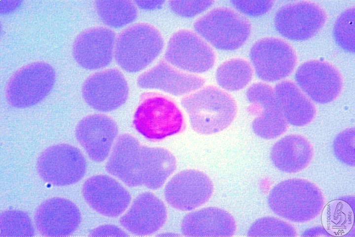
Lymphocyte: • Small and Globulous Cell • Nucleus with Dense Chromatin • Great Nucleus/Cytoplasm relation |
|
Lymphocyte
B Lymphocyte
T |
|
Plasmocyte: • Antibody-secreting cell • Characteristics of a secreting cell (well developed R.E.R. and G.A.) • Round and eccentric nucleus(dense and pale chromatin, radiated) |
|
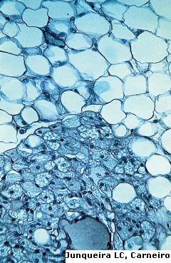
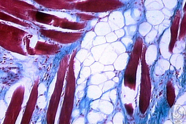
Adipocytes |
|
Functions: • Storage of lipids • Absorption of impacts • Heat insulator • Store liposoluble vitamins • Necessary for the synthesis of steroid hormones • All Cell Membranes need lipids • Can be use for the production of energy • Form the Panniculus, layer beneath the hypoderm • In optical microscopy, the negative image of the lipid is observed for it’s been dissolved by the alcohol. |
|
Unilocular
Adipocyte: • White fat • Present a single fat droplet • Flat and peripheral nucleus • Well developed S.E.R. • Supplies lipids to the multilocular adipocyte |
|
Multilocular
Adipocyte: • Brown fat • In mammals it’s present only in newborns(heat regulation) • Cytoplasm with a lot of fat droplets • Round nucleus • High concentration of mitochondria • All of the ATP produced in the cell is dissipated in the form of heat |
|
Undifferentiated
Mesenchymal Cells: • Differentiate into Connective Tissue cells (fibroblast and adipocyte) • Possess a shape very similar to a fibrocyte |
|
II - Extracellular Matrix Components: Collagen
Fibers: Types: I II III IV Ultra-structure: |
|
Elastic
Fibers: Elastogenesis: Oxytalanic Elauninic Elastic
Fiber Proper Types: Elastic Elauninic Oxytalanic Specific
Staining: |
|
III - Ground Substance Function: Formed of: Glycosaminoglycans
(GAGs) Proteoglycans Non-collagenous
glycoproteins • Examples: Fibronectin and Laminin, forms the Basal Lamina |
|