Imune
System |
Lymph
node
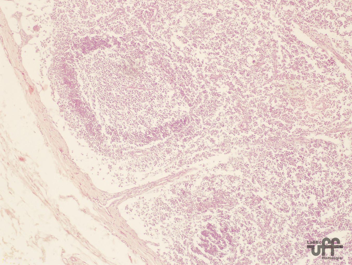
• It has a capsule and a kidney-like or round aspect
•
The capsule is formed by dense connective tissue that sends
septa into the node, dividing it into incomplete compartments.
• It presents a incurvature called hilum
• The parenchyma is divided into a cortical region, below
the capsule, and a medullar region, at the center of the organ
and the hilum.
• The region between the cortical and medullar region
is called the paracortical region and this is where we find
the T lymphocytes
|
|
Cortical
Region
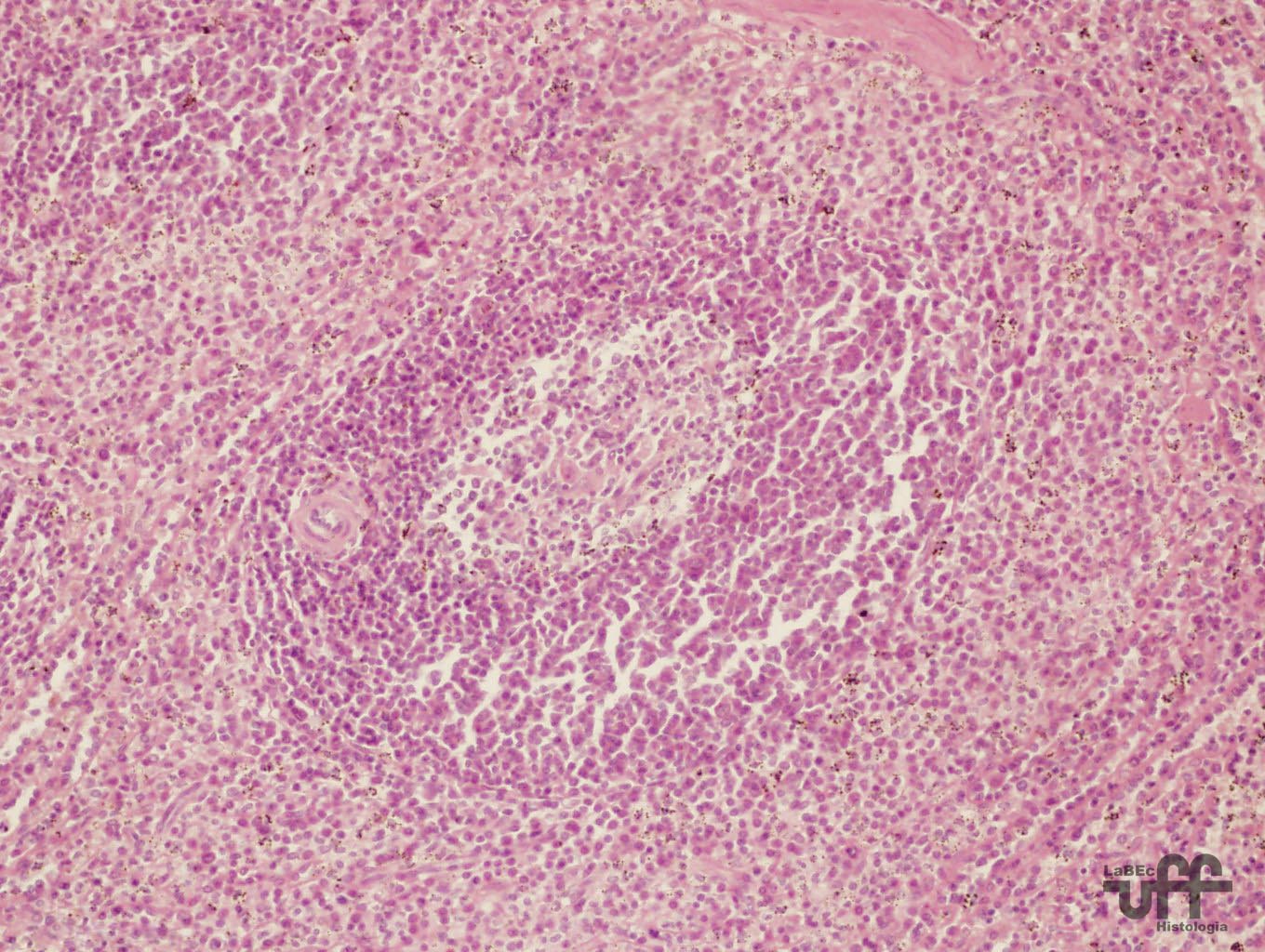
• Beneath the capsule and around the trabeculae we find
the loose lymphoid tissue forming the subcapsular and peritrabecular
sinuses
• The lymph runs inside the sub-capsular and peritrabecular
sinuses
• The loose lymphoid tissue presents predominantly reticular
cells, reticular fibers and fixed macrophages
• The
rest of the cortical region is formed by dense lymphoid tissue
with lymphatic nodules that can present a germinal center (central
region that is less stained)
• The dense lymphoid tissue is formed by reticular cells,
reticular fibers, fixed macrophages and mostly free cells(lymphocytes).
|
Lymphatic
Nodules
• Formed by reticular cells, reticular fibers, fixed macrophages
and free cells.
• The germinal center is formed by lymphoblasts, large,
medium and small lymphocytes, and plasmacytes in various developmental
stages.
Paracortical
Region
• Formed by dense lymphoid tissue that is populated by T
lymphocytes
|
|
Medullar
Region
• Presents medullary cords and medullary sinuses
• The medullary cords are formed by dense lymphoid tissue
• • The medullary sinuses are formed by loose lymphoid
tissue |
|
Spleen
• Presents a dense connective tissue capsule that presents
smooth muscle fibers.
•
From this capsule, septa are sent into the organ dividing it into
incomplete compartments.
• The parenchyma is divided into the White Pulp and the
Red Pulp.
|
 |
White
Pulp
• Formed by dense lymphatic tissue that forms a supporting
net of blood vessels.
• It possesses reticular fibers, mesenchymal reticular cells,
lymphoblasts and plasma cells.
• In some points of the parenchyma, a network of blood vessels
form nodules that contain, internally, an arteriole called the
central arteriole, however it is normally observed in an excentric
position in histological sections.
• The
structure represented by a nodule with an excentric arteriole
constitutes the characteristic elements for the histological diagnosis
of the organ, the Malpighian corpuscule.
• The dense lymphatic tissue is arranged in cords that are
called the Billroth cords.
|
 |
Red
Pulp
• Composed of loose lymphatic tissue that form splenic sinuses
that are set around the entire parenchyma surrounding the Malpighian
corpuscles and Billroth cords.
• There are also the sinusoid capillaries whose discontinuous
walls allow free passage of the blood which will occupy all of
the loose lymphatic tissue and therefore in anatomical sections
a redish color is observed.
• We also find the penicillate arteries, the reticular fibers
and characteristic cells of the loose lymphatic tissue
• It possesses reticular fibers, mesenchymal reticular cells,
lymphoblasts, lymphocytes and plasma cells
• The Billroth cords, though formed by dense lymphatic tissue,
can also be considered part of the red pulp due to its location.
|
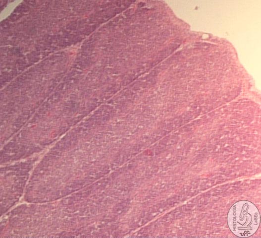
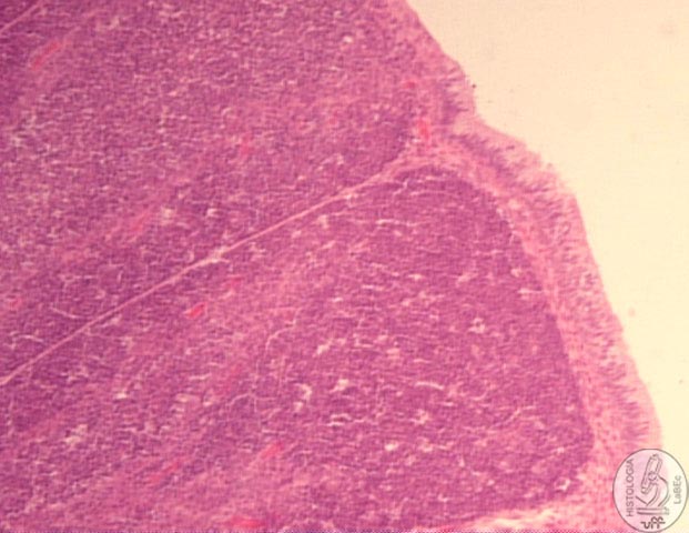
Thymus
• It has 2 lobes surrounded by a capsule of dense connective
tissue that emits septa dividing the organ into lobules, this
septation is incomplete, and therefore the separation of the lobules
is irregular.
•
The lobules present a cortical zone of dense lymphatic tissue
and a central zone( medullar) of diffuse lymphatic tissue with
mostly young cells (lymphoblasts)
• Lymphoid nodules cannot be found in the thymus, the cortical
and medullar region have the same types of cells, however in different
amounts, which are: the T lymphocytes in different phases of maturation
and reticular epithelial cells.
Cortical
Region
• Small lymphocytes predominate.
• Epithelial reticular cells are found in small amounts.
|
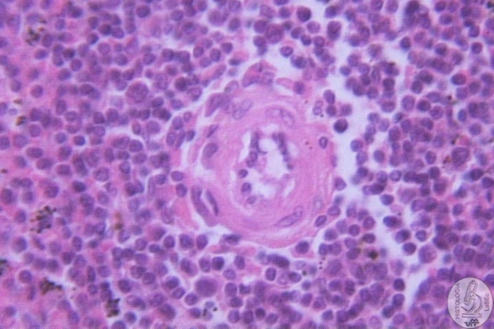 |
Medullar
Region
• We find lymphoblasts, young lymphocytes and epithelial
reticular cells.
• We also observe epithelial reticular cells in concentric
layers, forming the corpuscles of Hassal.
• We can find at the center of the Hassal Corpuscles, cell
debris that are many times calcified
|
|
Bursa
of Fabricius
• It is a dorsal saccular diverticulum of the proctodeum,
exclusive to birds.
• Characterized by tall and thick mucous folds (plicae)
filled with numerous polyhedral follicles
• Each follicle, which is composed of lymphatic tissue,
is divided into a cortex and medulla.
• A layer of undifferentiated epithelial cells occupies
the periphery of the medulla that is separated from the cortex
by a capillary layer.
• The
bursa is lined by pseudo-stratified cylindrical epithelium, except
at the apex of each follicle where it is lined by simple cylindrical
epithelium.
|
 |
|
 |
|