| Veterinary
Histology UFF Department of Morphology - Biomedic Institute LaBEc - Laboratory of Cellular and Extracellular Biomorphology |
|||
Veterinary
Histology Atlas |
|||
Epithelial
Tissue |
|||
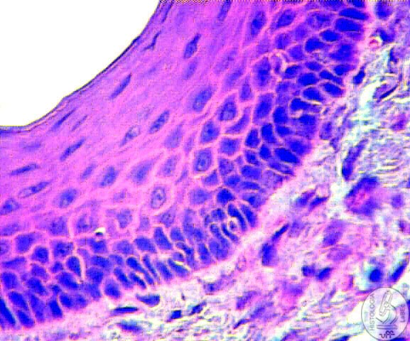
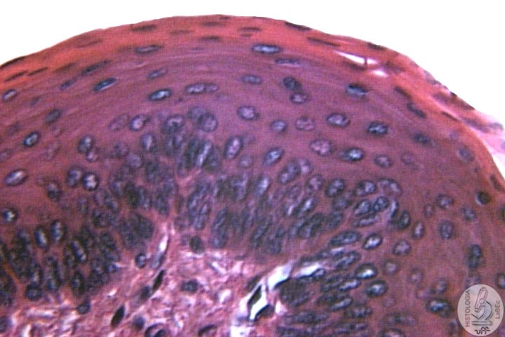
General Characteristics •
Polyhedral Shape |
|
||
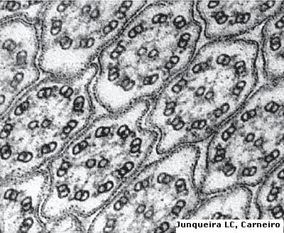
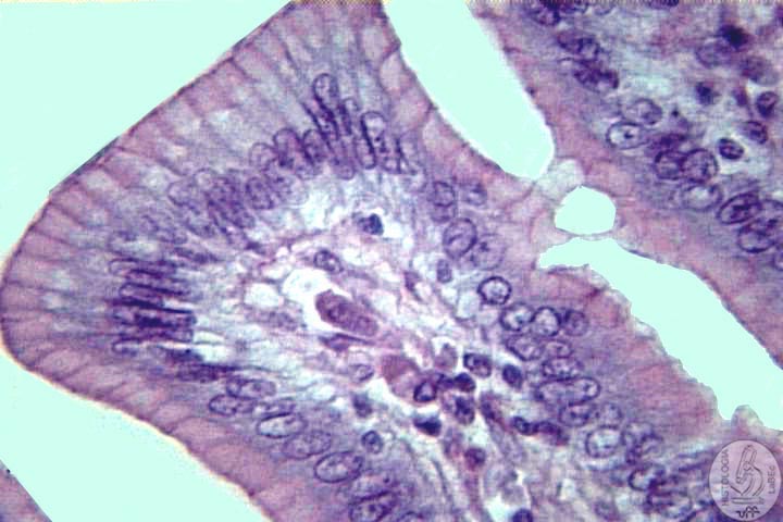
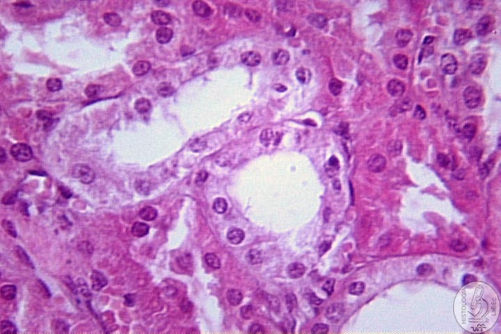
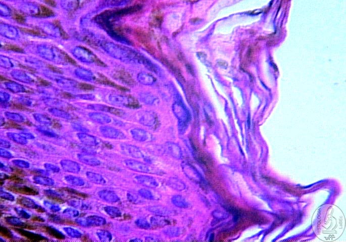
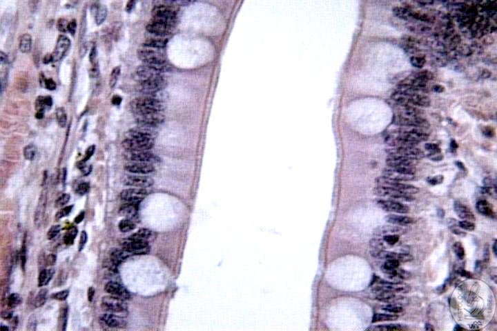
Membrane Specializations - Apical Pole Microvilli: |
|||
|
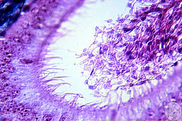
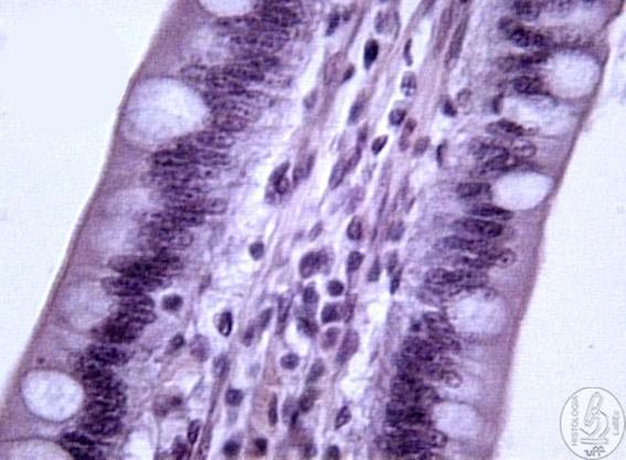
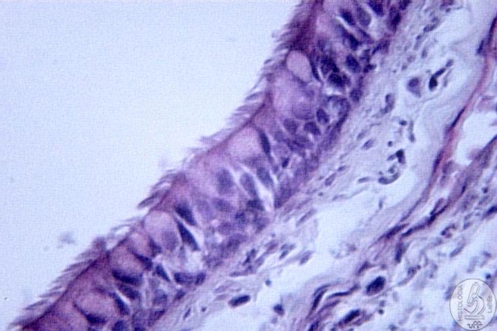
Cilia: |
||
|
Estereocilia:
|
||
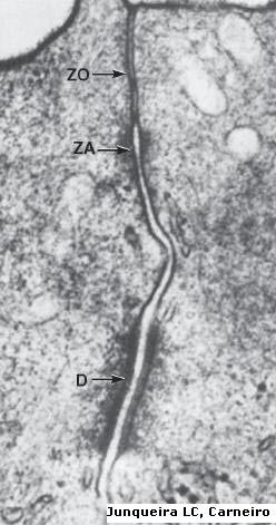
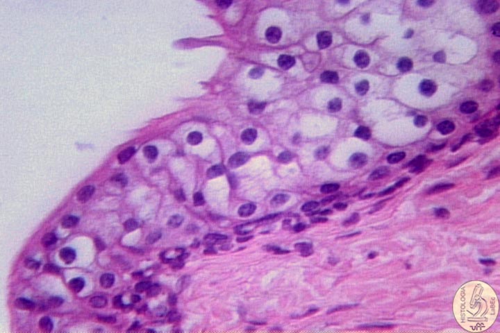
Basal
Lamina |
|||
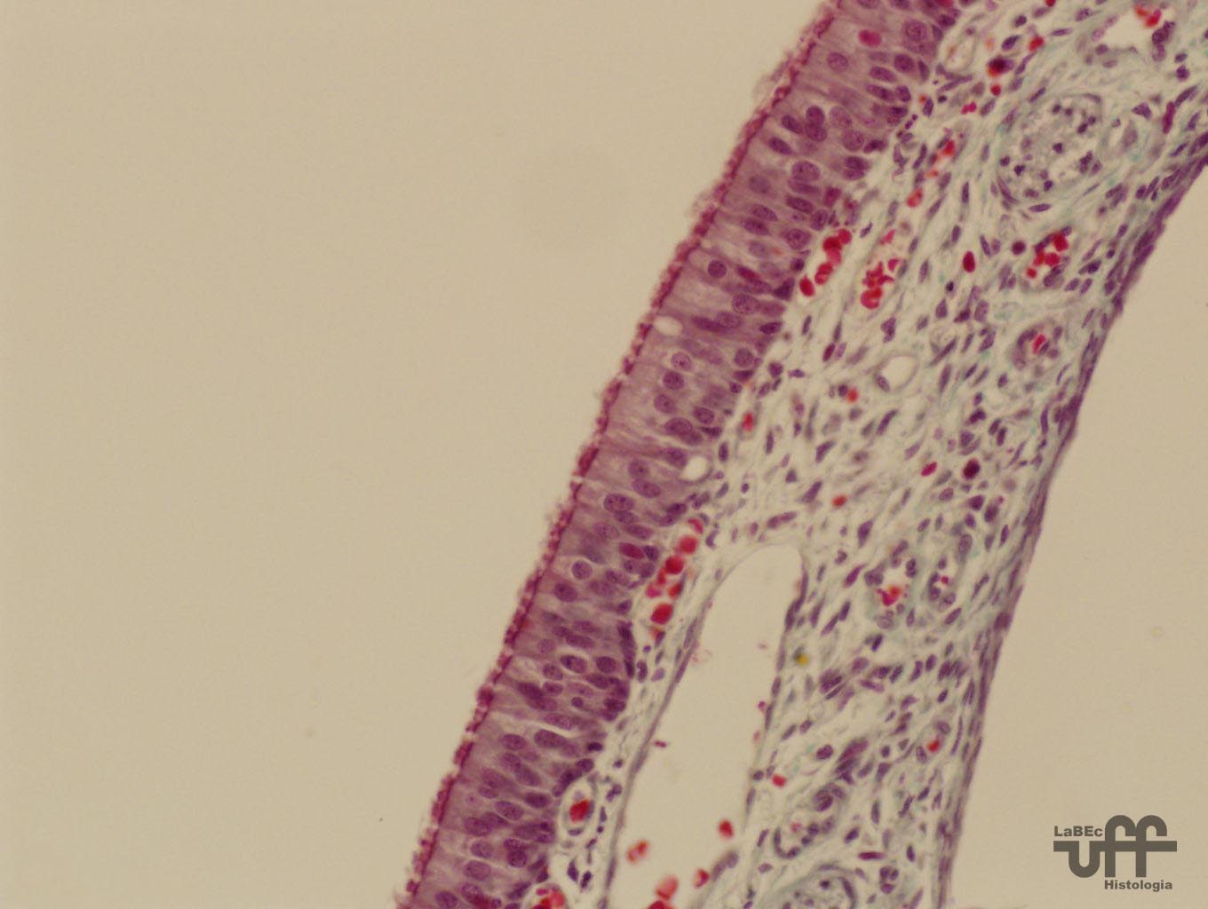
Lining
Epithelial Tissue Classification: According to number of Stratums: |
|||
|
|
|
|
| • Simple: All cells are in contact with the basal lamina | • Stratified: At least one cell does not touch the basal lamina | ||
|
|
|
|
| • Pseudostratified: All cells are in contact with the basal lamina, but they have variably placed nuclei. | |||
According to the Shape of the Cell: The shape is always given by the more SUPERFICIAL layers. |
|||
 |
 |
|
 |
| • Squamous | • Cuboidal | •
Columnar or Prismatic |
•
Globulous |
According
to membrane specializations and Cells or Accessories : |
|||
 |
 |
 |
 |
| • Keratin | • Goblet Cells | • Striated border | • Cilia |
Glandular Epithelial Tissue Group of Epithelial Cells that associate themselves and differentiate cellularly, being capable of producing and secreting substances. Exocrine: • Releases its secretion through ducts to the outside • Composition: - Glandular Portion (adenomere) - Conducting Portion (duct) Classification: According to Excretion Mechanism : • Holocrine - Cell disintegrates along with its excreted content - Stem-cells originate new secreting cells - Example: Sebaceous Gland • Merocrine - There is no alteration in the cell’s morphology - Cytoplasmic granules fuse themselves with the cell membrane and release their contents - Example: Sweat Gland • Apocrine - Loss of the cell’s apical portion when excretion is released - Example: Mammary Gland According to the Morphology of the Secreting Portion: • Acinar • Tubular • Alveolar According to the Conducting Portion: • Simple: One duct • Compound or Branched: More than one duct |
|||
According to the Quality of the Secretion: |
|||
 |
 |
 |
|
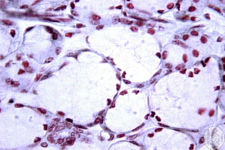
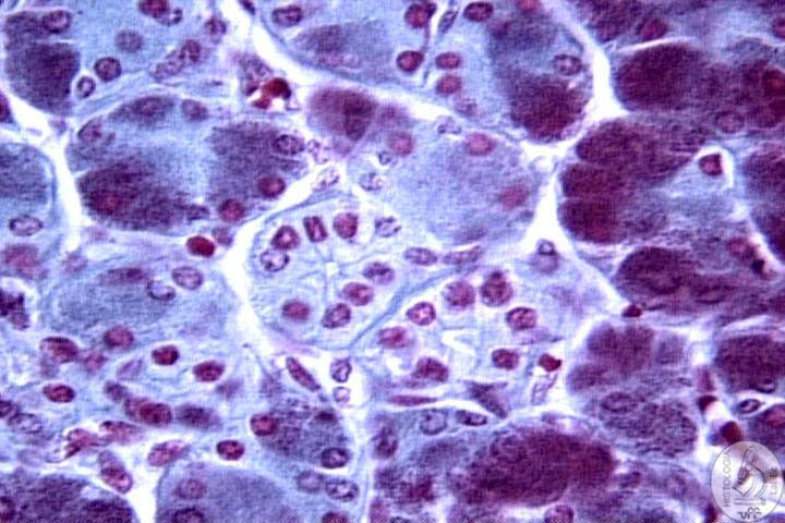
•
Mucous: Glycoproteic, viscous, flat nucleus |
•
Serous: Proteic, liquid, round nucleus |
•
Mixed: Demilunes serous on mucous acinus |
|
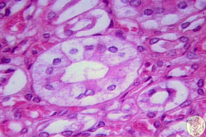
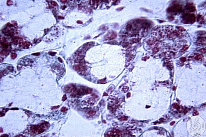
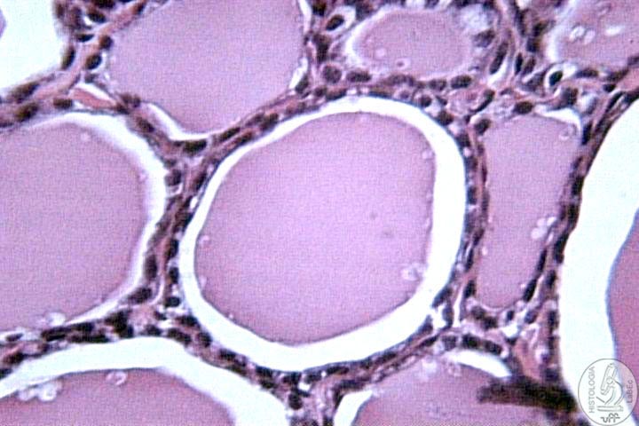
Endocrine: • Releases its secretion directly into the connective tissue • Morphology: - Cordonal ( cordon of cells, no deposit) |
|||
-
Follicular (in the shape of a hollow “ball”, possess
a colloidal deposit) |
 |
||
Specialized: |
|||