| Veterinary
Histology UFF Department of Morphology - Biomedic Institute LaBEc - Laboratory of Cellular and Extracellular Biomorphology |
|||
Veterinary
Histology Atlas |
|||
Muscle
Tissue |
|
General Characteristics •
Mesodermal Origin |
|
| Types | |
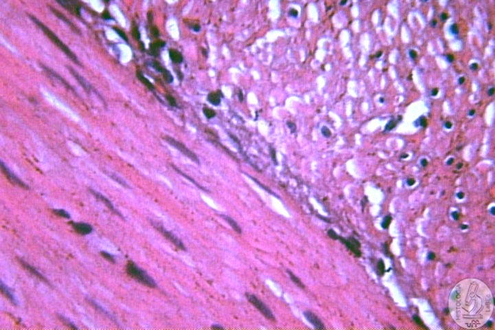
Smooth Muscle •
Long Spindle-shaped Cells |
|
•
Mononuclear cells, central nucleus |
|
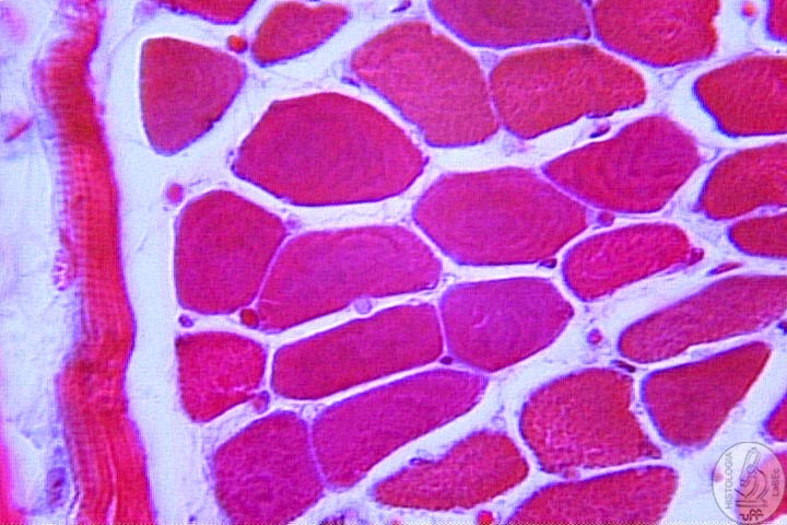
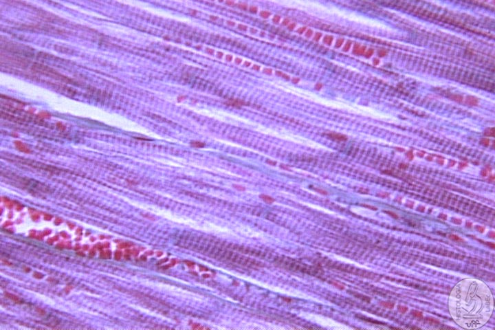
Striated skeletal muscle •
Elongated cylindrical cells |
|
•
Present transverse striations |
|
Cell Structure Myofibrils:
Formed by thin and thick filaments Sarcomere:
morphofunctional unit of striated muscle fibers Sarcoplasmic Reticulum: Regulates specifically the flow of calcium ions Transverse
tubules (T system): |
|
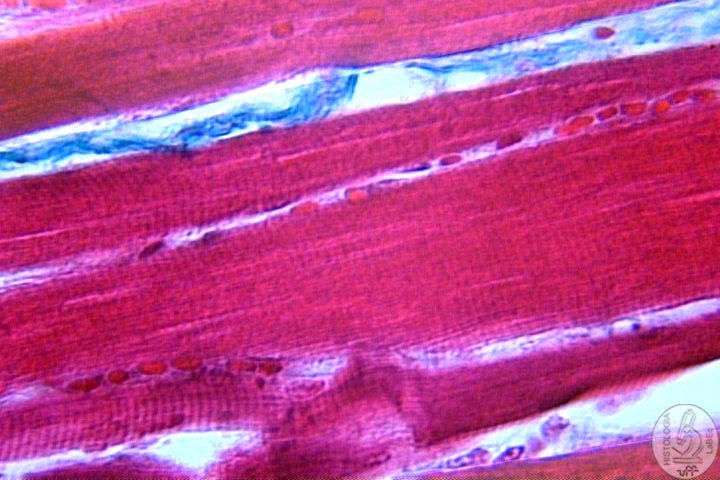
Striated Cardiac Muscle •
Formed by elongated cells that anastomose themselves |
|
Muscular Contraction •
The motor end plate is where the nerve inserts itself in the muscle
fiber |
|