| Veterinary
Histology UFF Department of Morphology - Biomedic Institute LaBEc - Laboratory of Cellular and Extracellular Biomorphology |
|||
Veterinary
Histology Atlas |
|||
Circulatory
System |
|
Comprises
the cardiovascular and lymphatic vascular system |
|
Cardiovascular System Heart |
|
• It’s the pump of the cardiovascular system |
|
| •
Formed by four chambers: -Atria(2) – receive blood -Ventricles(2) – send blood • Possesses three layers: |
|
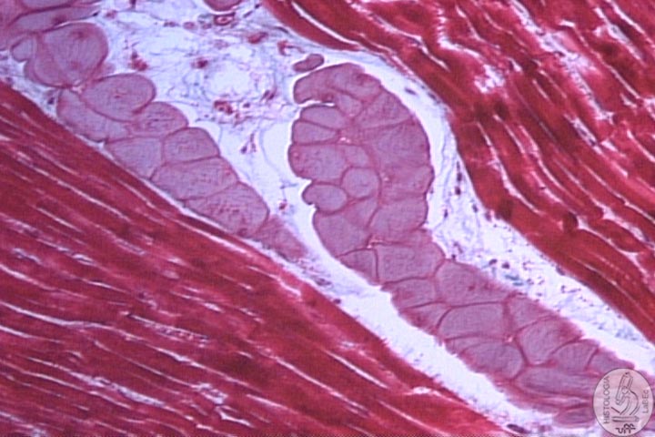
| I
- Endocardium • Internal lining of the heart • Presents endothelial cells(simple squamous epithelium) and loose connective tissue • Beneath the endocardium, we find the Purkinje fibers |
|
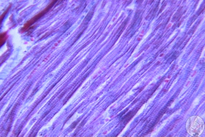
II
- Myocardium III
- Epicardium |
|
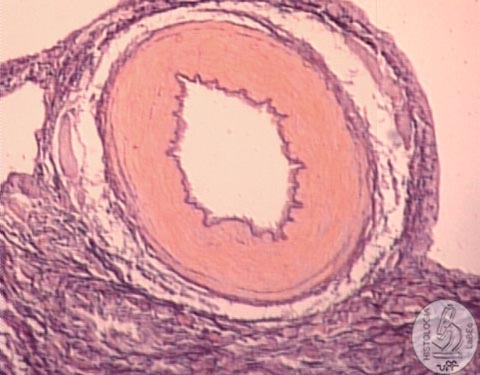
| General Structure of Vessels | |
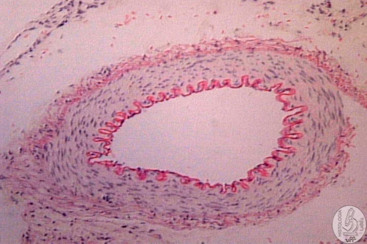
| Tunica
Intima • Layer of endothelial cells that line the lumen of the vessel • Layer of loose connective tissue • Internal elastic lamina (separates the tunica intima from the media) |
|
Tunica
Media Tunica
Adventitia |
|
Arteries •
Transport blood from the heart to the capillary beds |
|
| - Elastic arteries – conducting | |
| - Muscular arteries – distributing | |
| - Arterioles – supply blood to the capillary networks | |
| - Metarterioles - form the capillaries | |
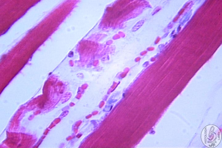
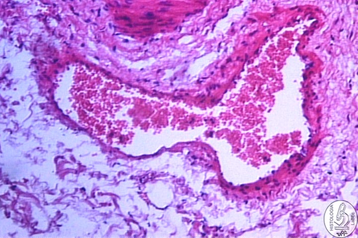
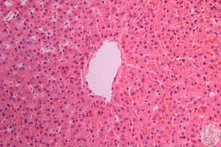
Capillaries •
Formed by a single layer of endothelial cells |
|
| - Continuous Capillaries: Do not have interruptions in their walls | |
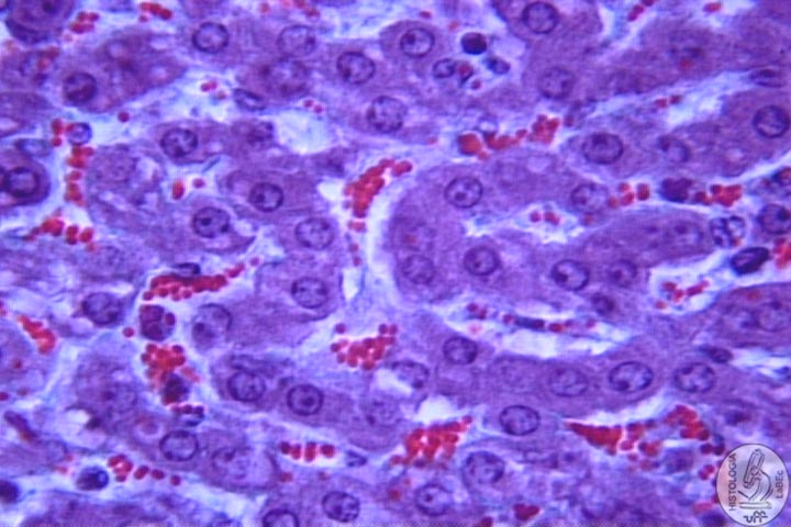
| - Sinusoidal Capillaries: Irregular, adapt themselves to the shape of the structure in which they are located | |
| - Fenestrated Capillaries: Present pores or fenestrae in their walls | |
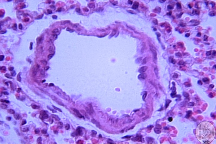
Veins •
Transport blood from the capillary beds to the heart |
|
 |
- Large Veins |
- Small Veins |
|
- Venulles |
|
| •
Along the capillaries and veins we find pericytes(contractile function) • Vaso vasorum -Are the arterioles, capillaries and venules -Responsible for the nutrition of the tunica media and adventitia of large vessels(thick walls) |
|
Lymphatic Vascular System •
Vessels that remove the excess tissular fluids in the interstitial
spaces |
|