Veterinary Histology UFF
Department of Morphology - Biomedic Institute
LaBEc - Laboratory of Cellular and Extracellular Biomorphology
 
Veterinary Histology Atlas
    Versão em Português
Introduction
Urinary System of Birds

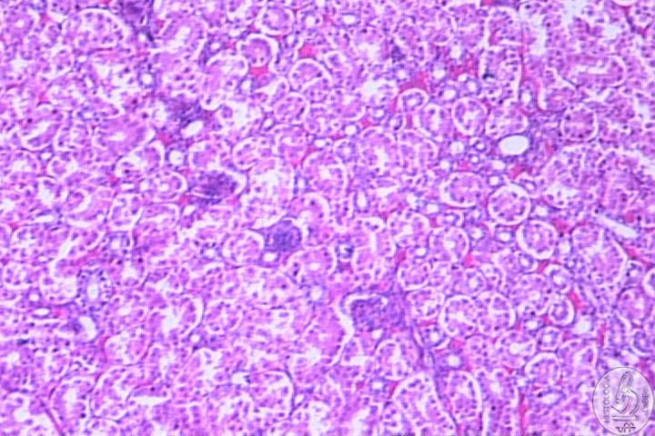
The urinary system of chickens consists of a pair of large elongated kidneys. The ureters drain each kidney and open into the urodeum of the cloaca. There is no renal pelvis or bladder in birds.

Components

Kidney

Subdivisions of each kidney
Cranial
• Intermediate
• Caudal

Each subdivision is composed of lobules
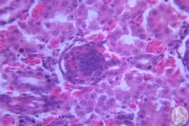
Each lobule consists of a large cortical component and a smaller medullar component.
• All the lobules that drain into a single branch of the ureter form the lobe.


There are two types of nephrons
• The cortical type
- is more numerous
- does not possess a Henle loop
- It is located entirely inside the cortex.
- It has a smaller renal corpuscle than the medullar nephron.
• The medullar type
- is less numerous
- It has a Henle loop that extends itself from inside the medulla.


• Each glomerulus contains a compact mass of mesangial cells at its center.
A layer of podocytes with large round nuclei cover the surface of the glomerular capillaries forming the glomerular epithelium of the Bowman capsule.
• The capsular (parietal) layer of Bowman’s capsule consists of a simple squamous epithelium.
• Juxtavascular cells and a macula densa associate themselves with the renal corpuscle at its vascular pole.

Collecting Tubules

• The collecting tubules occur in the more peripheral walls of the cortex.
• They are lined by pale cells with cuboidal or columnar shapes.
• The collecting tubules join the distal contorted tubules to the perilobular collecting ducts.

- The latter join with those of the other lobules to form the medullary tracts.
- The tracts group to form a medullary cone.
- Each cone ends in a single branch of the ureter.
• The lining of the smaller collecting ducts is simple cuboidal epithelium. It gradually becomes simple columnar and finally turns into pseudostratified columnar close the ureteral branch.

Ureter

• The ureter of chickens is a muscular tubule of approximately 2mm in diameter.
• Its wall consists of mucosa, muscular and adventitial layers.

Mucosa
• It is generally lined by a pseudostratified columnar epithelium.
• Underneath the epithelium, there is a thick layer of loose connective tissue that contains various amounts of diffuse lymphatic tissue and sometimes a lymph nodule.

Muscular
• Each muscle layer consists of an internal longitudinal layer and an external circular smooth muscle layer.
• A third external longitudinal layer is found close to the cloaca.

Adventitia
• The adventitial layer consists of a layer of loose connective tissue.

Credits
Tissues
Epithelial
Connective
Cartilaginous
Osseous
Nervous
Muscle
Systems
Circulatory
Blood
Imune
Digestive
Accessory Gls.
Respiratory
Integumentary
Urinary
Endocrine
Male Reprod.
Female Reprod.
Birds Histology
Quiz
Videos
Bibliography