| Veterinary
Histology UFF Department of Morphology - Biomedic Institute LaBEc - Laboratory of Cellular and Extracellular Biomorphology |
|||
Veterinary
Histology Atlas |
|||
Respiratory
System of Birds |
|||||||
| The respiratory system of birds is formed by nostrils, a nasal cavity, a pharynx, a trachea, a syrinx, bronchi, air capillaries and air-sacs. •
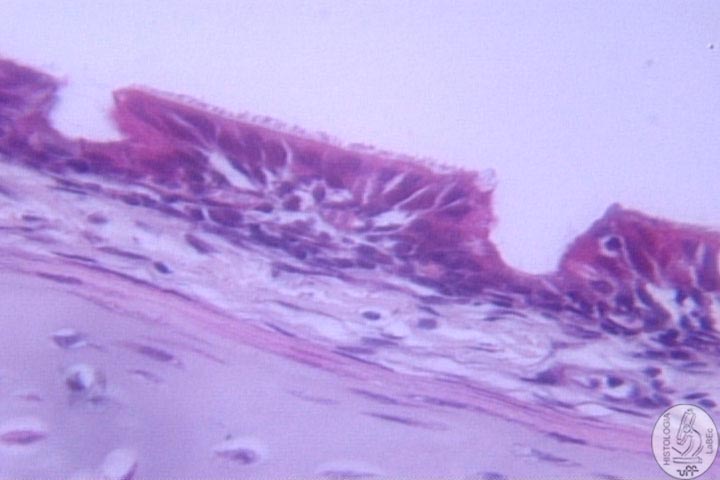
The skin enters the nostrils up until the first part of the
nasal cavity (the vestibule), that is lined by modified keratinized
stratified squamous epithelium. It is characterized by epithelial
cells organized into columns, giving it a ceruminous aspect
on the surface. Pharynx
• The pharynx is lined by a stratified squamous epithelium.
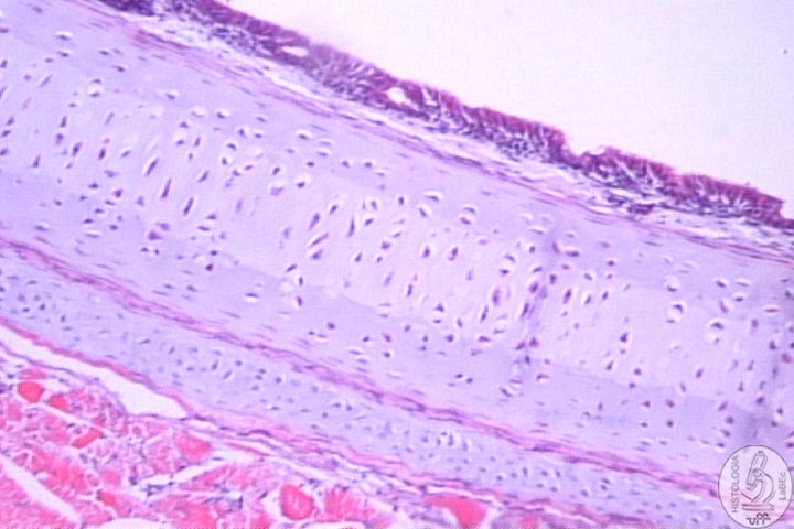
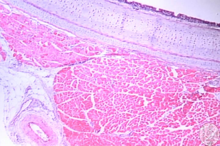
Trachea
• At the anterior extremity of the trachea there is a
cranial larynx that is reinforced by a cartilage ring. |
|||||||
| |
•
It is lined by a ciliated pseudostratified columnar epithelium
that contains numerous simple mucous alveolar glands. |
||||||
Syrinx
• The syrinx or voice-box is located in the thoracic cavity
, at the point of bifurcation of the trachea into two bronchi.
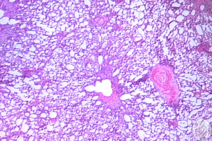
Bronchi
Each extrapulmonary primary bronchus enters the lung as
a primary intrapulmonary bronchus (mesobronchus). Primary
bronchi Secondary
bronchi Tertiary
bronchus
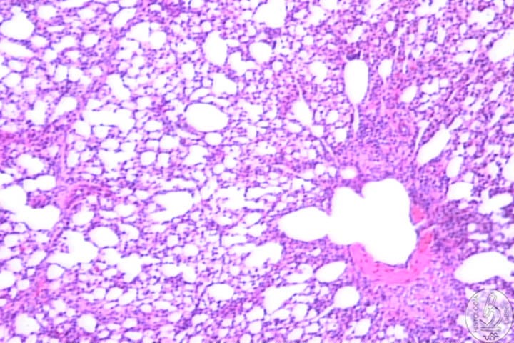
Air-sacs
The air sacs are thin-walled, paired or not structures
that occur in the cervical, clavicular, thoracic and abdominal
regions of the body.
• The air sacs are lined by squamous columnar, ciliated
cuboidal and ciliated columnar cells. The sacs are poorly vascularized and do not participate in the gas exchange.
|
|||||||